Focus Areas
Research at MRRI has an international reputation for excellence. Our work is primarily conducted in three focus areas:
Movement Science
Movement control is one of the most complicated tasks our nervous system has to manage. The human body has over 600 muscles, whose activity must be coordinated to control the movement of over 200 bones. Daily tasks like folding laundry, standing up from a chair, walking to the kitchen, and pouring a glass of water can seem effortless. In reality, they require the nervous system to perform many computations. Movement impairments that may result from nervous system damage can cause substantial disability and impact quality of life. Research in this Focus Area aims to understand how the nervous system controls, learns, and re-learns movements and how damage and recovery of specific nervous system structures impacts these processes. This knowledge is critical to design evidence-based rehabilitation protocols for people with movement impairments, including individuals with stroke, Parkinson’s disease, cerebellar damage, and spinal cord injury. Our research spans basic science investigating the processes involved in motor control and motor recovery through the development and evaluation of interventions to enhance motor recovery and treat movement impairments in clinical trials.
Cognition, Behavior, and Emotion
How we think, feel, and act is what makes us who we are. Cognition, behavior, and emotion arise from an intricate interplay of multiple brain regions working together. This Focus Area encompasses our interests in numerous domains of higher-order human functioning including attention, cognitive control, memory, spatial processing, emotional processing, and skilled action. Our work examines the brain regions and systems that are critical for cognition, behavior, and emotion, as well as how function is influenced by neurological conditions like stroke, traumatic brain injury (TBI), and Parkinson’s disease. We also develop and evaluate theory-driven assessments and treatments that target performance in these areas. One area of particular focus is the executive system, a pivotal regulator across many domains, including decision-making, problem-solving, emotional regulation, motivation, reward processing, and goal-directed behavior. Dysregulation in the executive system often impacts people with neurological conditions, manifesting as cognitive deficits, difficulties with behavioral regulation, and emotional challenges. Our work examines how the executive system controls language, motor, and other cognitive functions, and how social and contextual factors interact with cognition, behavior, and emotion, contributing to functional behaviors.
Language and Communication
Human communication is a multifaceted process that draws on language, cognitive, and sensory-motor skills, and is fundamental to most aspects of human activity. This Focus Area brings together researchers who study the mechanisms, neuroanatomy, and functional aspects of language and communication. Areas of interest include language production, language comprehension, multimodal communication (verbal, facial expressions, gestures, etc.), and functional communication, which takes into account social and environmental contexts and the goals of all conversational partners. Stroke, brain injury, and other neurological disorders often affect language and communication abilities by causing aphasia—a disorder of language—or limb apraxia, a deficit in action planning that affects the ability to produce and understand gestures. Our research seeks to advance theoretical models of language and communication processing in people with and without aphasia and apraxia. A main theme involves translating from basic research on language processing and communication to inform therapeutic approaches for addressing aphasia and apraxia. Research designs span the translational pipeline, from basic, pre-clinical studies to establishing efficacy of different treatment approaches to treatment implementation in the clinic.
MRRI Core Facilities
Scientists at MRRI have expertise in a broad array of methodological approaches used in neurorehabilitation research. MRRI’s infrastructure supports the recruitment of research participants, and our Methodological Cores compile and maintain the tools, resources, and procedures that are used across MRRI’s 10 research laboratories and programs.
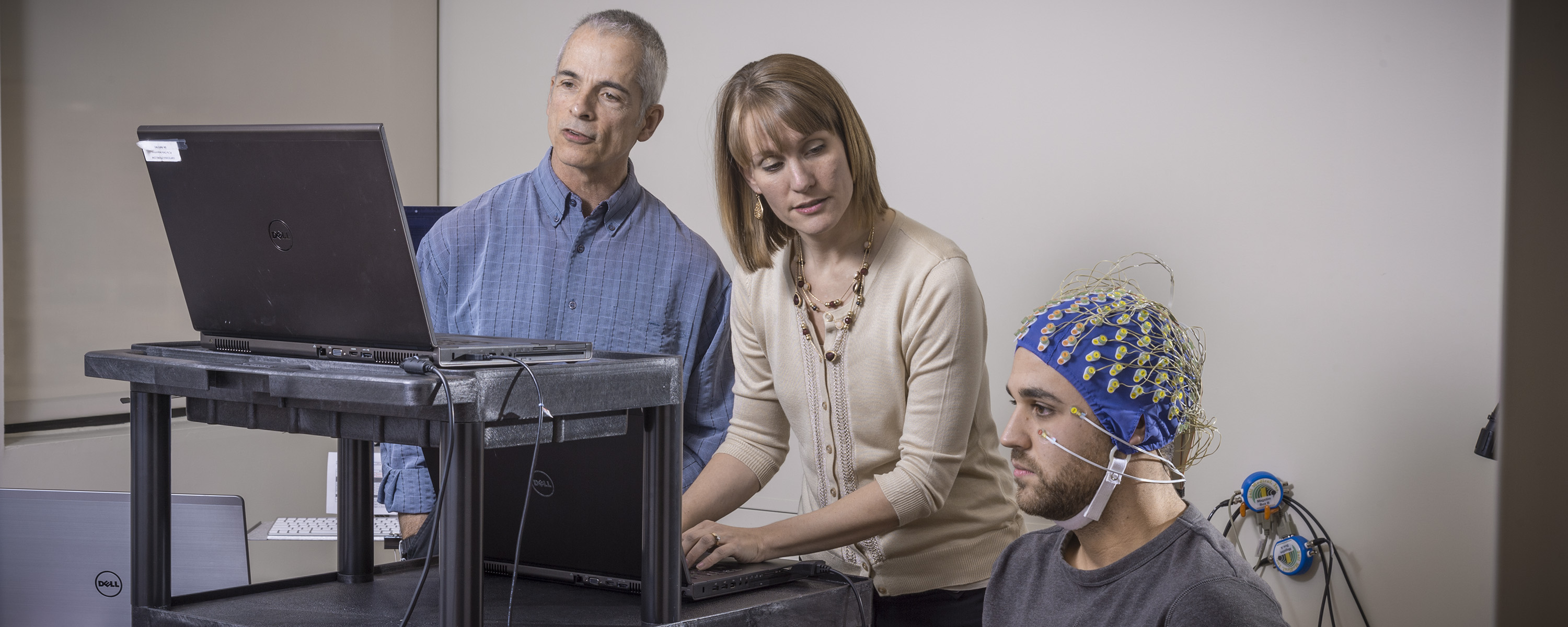
Brain Structure & Function Core:
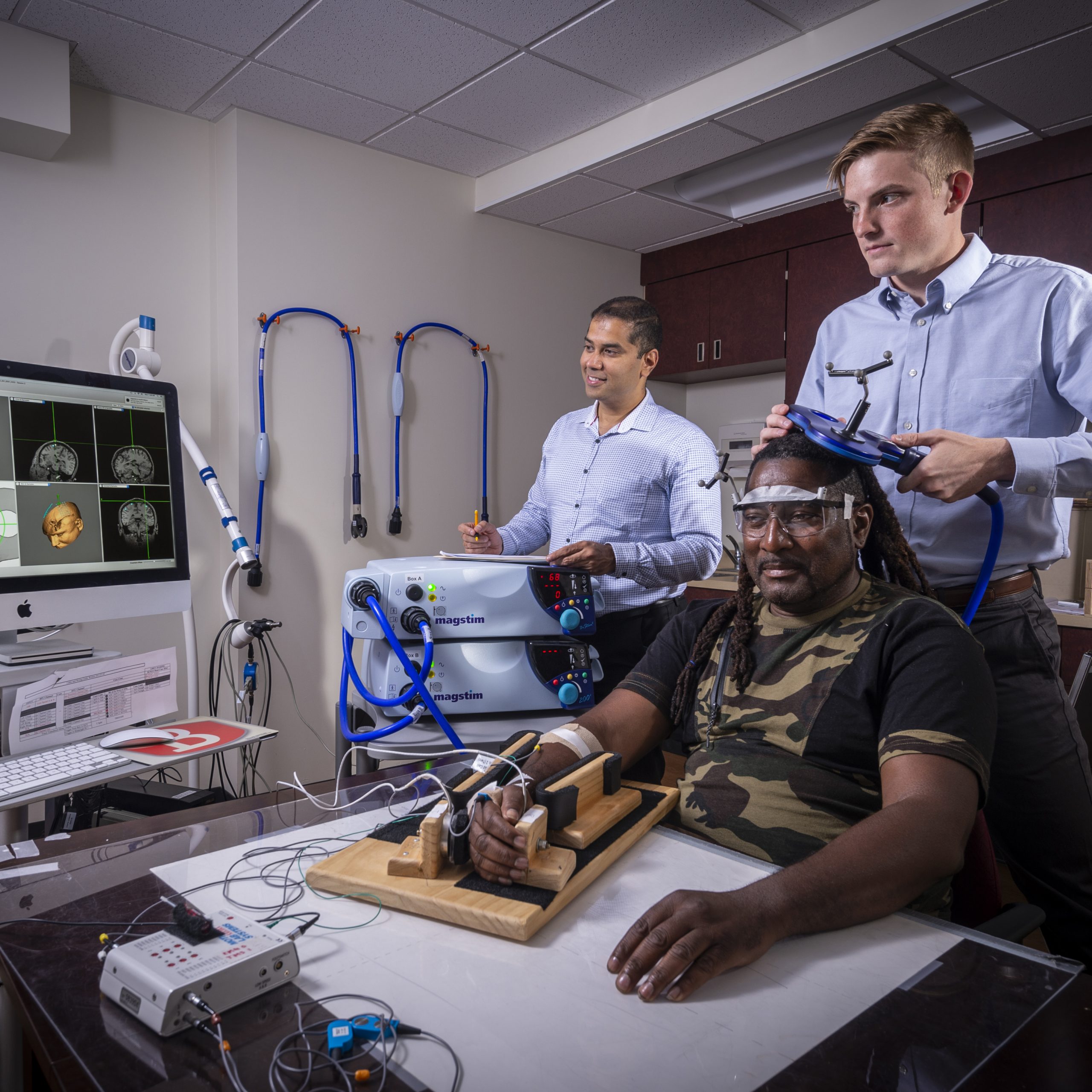
The Brain Structure and Function Core provides centralized resources, protocols, technical information, and methods for neuroimaging (including structural and functional magnetic resonance imaging), electrical stimulation, and electroencephalography across MRRI labs to optimize organization and efficiency. Current documentation includes:
- A database tracking progress in obtaining imaging and lesion segmentation of MRRI research participants
- Detailed scanning protocols across Penn and Jefferson sites
- Lesion mask files
Measurement Methodological Core:
The Measurement core maintains a shared resource library that documents assessments and assessment resources (e.g., databanks such as AphasiaBank) across content areas. Available documentation includes:
- Domain(s)/sub-domains assessed
- Appropriate population(s)
- Standardization and psychometric information
- Published benchmarks for change
- Instrument availability
Motion Analysis Core: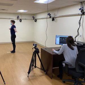
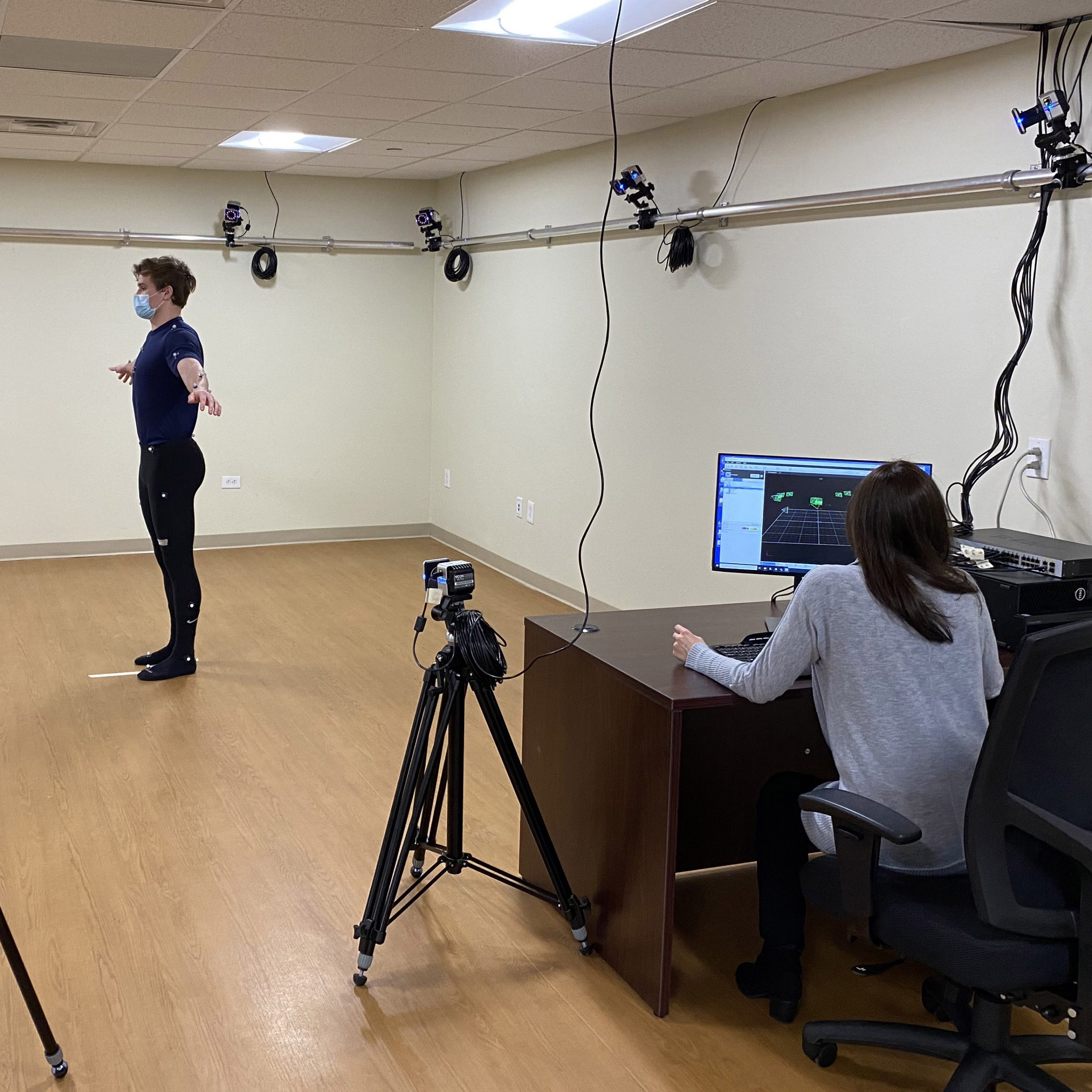
The Motion Analysis Core oversees specialized equipment shared across MRRI laboratories, used in studies to understand how the nervous system controls and learns movements and how damage to specific brain structures impairs these processes. These resources include:
- Two systems for precise 3-dimensional optical motion capture (Codamotion, Vicon) and a system for magnetic motion capture (NDI trakSTAR) for cases where optical tracking is unsuitable.
- Two virtual reality devices (Oculus Rift S, Oculus Quest 2) that can be integrated with optical or magnetic motion capture using custom-developed software, which permits the study of visual integration in movement control.
- Two robotic devices (KINARM Bimanual Exoskeleton, ARMEO Power) to allow additional investigation of somatosensory and haptic influences on movement control.
- Eye tracking systems (Eyelink 1000, Pupil Labs) to monitor gaze position while individuals are viewing stimuli and generating movement or to study the control of eye movements in their own right.
- Three electromyography systems: two wired (CED, Motion Labs) and one wireless (Delsys Trigno) which can be used to simultaneously record muscle activation while tasks are performed using the motion capture or robotic equipment.
MRRI Virtual Reality Core: 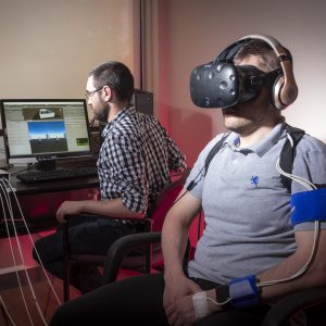
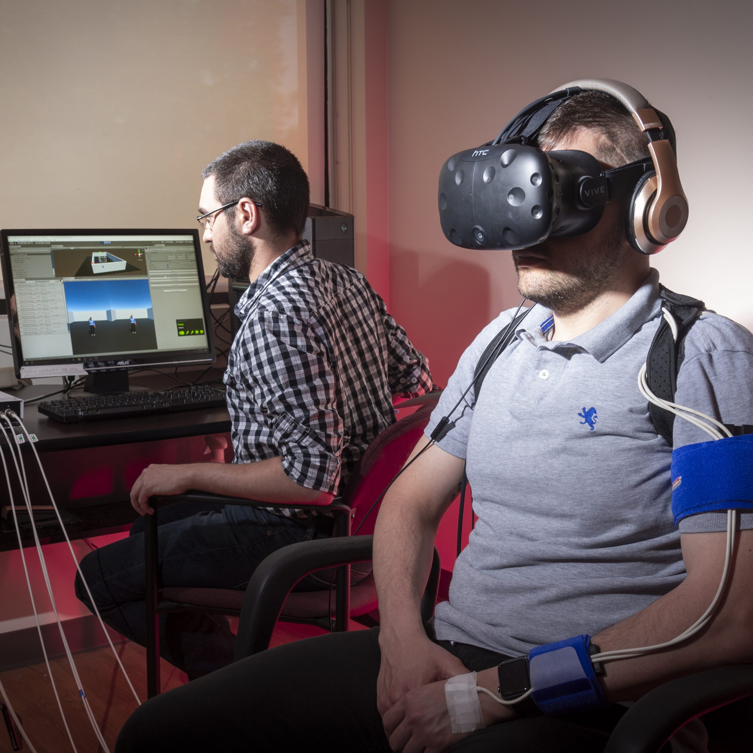
This shared facility provides the infrastructure to support experiments conducted using virtual reality. Available equipment includes:
- two head-mounted virtual-reality systems (Vive, HTC and Oculus Quest, Oculus) each with two hand-held controllers and associated optical tracking system.
- a magnetic tracking system (TrakSTAR, Ascension Technologies).
- an optical hand-tracking system (Leap Motion.
- an integrated eye-tracking system (Pupil Labs).
- a dedicated PC running custom-written software in Unity.