Using Gestures and Brain Stimulation to Enhance Language in People with Stroke

What’s the word? Those tip-of-the-tongue moments, when a word is seemingly right there but just out of reach, can temporarily hinder our ability to communicate effectively. Everyone experiences word-finding challenges (called anomia) from time to time. However, for people who have aphasia, an acquired language disorder following stroke or brain injury, these word-finding challenges can be very frequent and disruptive, causing problems for everyday communication needs. Verbs present a particular challenge for the majority of people with aphasia. Verbs are vital because we need them to communicate about actions and events, and they are central to sentence structure and conveying relationships between subjects and objects. Without using verbs, language can sound telegraphic or robotic, and it becomes very hard to get a point across.

Gestures can help people retrieve words when they get stuck. Using a gesture to pantomime an action can convey a speaker’s intended message to others, and it can furthermore help the person produce the verb they want to say. Unfortunately, producing gestures can also be impaired following stroke, and many patients who have aphasia also have limb apraxia, a disorder that impairs skilled action and gesture production.

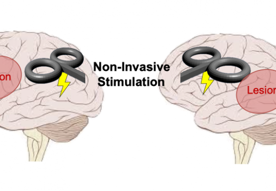
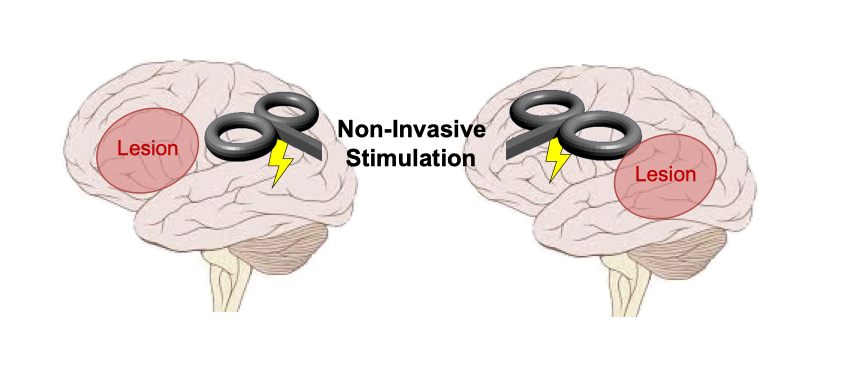
However, it is possible that simply watching someone else produce a gesture may help adults living with stroke to get those words off the tip of their tongues. This is possible when an observed gesture and intended verb share meaning, or semantics. Semantic knowledge consists of everything we know about the world, and it is stored in long-term memory. Semantics are distributed in a network of regions across the brain and can therefore be resilient to damage. Neurorehabilitation treatments that enhance semantic activation may therefore benefit many patients with different patterns of brain damage. Moss Rehabilitation Research Institute (MRRI) and University of Pennsylvania researchers Haley Dresang, PhD, Laurel Buxbaum, PsyD, and Roy Hamilton, MD, MS, are combining gesture observation with non-invasive brain stimulation to investigate whether enhanced activation of action knowledge (semantics) can facilitate verb production in patients with aphasia. This is the first study to implement a specific type of excitatory stimulation – intermittent theta-burst stimulation – on verb-production impairments, and also the first to combine brain stimulation with gesture-observation cues in aphasia. First, this research examines whether passive observation of gestures can help patients produce verbs. Second, this research compares the benefit of gestures on verb production in patients who have had a stroke that damaged the left anterior versus the left posterior parts of the brain. Third, Dr. Dresang and colleagues are also investigating whether applying non-invasive brain stimulation to increase activation in intact nodes of the semantic neural network will enhance the benefits that observing gestures may have on verb production. The researchers will apply a magnetic current that amplifies the activity of neurons in brain regions important for semantic processing, and they will assess whether patients are able to successfully name more verbs based on observed gestures while receiving this brain stimulation. This study will begin recruiting participants later this summer. This research advances scientific understanding of how the brain functions following injury. Furthermore, this work seeks to create a novel neurorehabilitation approach that will improve treatment success for a variety of stroke patients with language and/or motor impairments.


Understanding Spatial Navigation Challenges After Stroke

Erica Barhorst-Cates Headshot

To find the offices at Moss Rehabilitation Research Institute (MRRI), take the elevator to thethird floor. Once you exit the elevator, turn left, then right down the first hallway, then left through the conference room. Turn right, then left, and the office is the third door on your right. Got that? Okay, now head back to the elevator the way you came. Make sure not to bump into any of the obstacles in your way, and make sure you still pay attention to what the research assistant is saying as you go.

Spatial navigation, or the way we find our way around an indoor or outdoor space while remembering important landmarks or routes, is a complicated activity that many of us take for granted. It requires attention, memory, language, and, often, physical requirements such as the ability to walk or control a wheelchair. Spatial navigation involves various brain regions, including the temporal and parietal lobes. Even many neurotypical individuals have difficulty with navigation; some of us forget where the exit is in the local Target or have no idea which way is north.

Stroke can make the act of spatial navigation even more challenging, because lesions in different parts of the brain can impair different aspects of navigation. MRRI researchers Drs. Erica Barhorst-Cates, PhD, Aaron Wong, PhD, and Laurel Buxbaum, PsyD, are conducting research to better understand how stroke can affect navigation abilities, and in what situations. While one individual might have an excellent “mental map” of the environment, she may have difficulty walking and become distracted by the need to avoid an obstacle in the environment. This distraction may make her temporarily forget where she is, thus affecting her navigation ability. Another individual may walk normally but have impaired working memory ability, thus making it hard for him to remember a list of directions. Depending on where an individual’s stroke occurred in the brain, different abilities might be affected. There is no one-size-fits-all navigation impairment.

In their ongoing study funded by the Albert Einstein Society, Dr. Barhorst-Cates and colleagues are asking participants with stroke and a group of neurotypical participants to navigate around the MRRI building and learn the location of “landmarks” in the hallways. Much of the prior research on navigation after stroke has used computer based virtual reality tasks, where participants are seated and stationary. The task used in this study is more similar to how navigation occurs in the real world, thus allowing the researchers to more accurately understand the various challenges. They are testing participants’ memory for the object locations and the traveled routes using a series of measures that they have adapted for use for individuals with stroke. In addition to this navigation task, participants also complete a series of tests aimed at understanding their physical and mental abilities. The researchers have currently run 30 participants through the study out of our goal sample size of 55. We hope to use the results from these tests to understand more about which types of patients might be affected in terms of their navigation abilities.

Ultimately, the results from this study may help inform development of therapies to improve navigation abilities for individuals who suffer a stroke. In the meantime, which way is south?


Interview with Dr. Shailesh Kantak on the People Behind the Scientist Podcast

Dr. Kantak's headshot

In a new episode of the People Behind the Science podcast, Shailesh Kantak, PT, PhD, discusses his career, ongoing research on bimanual coordination after stroke, and his interests outside of science. Over the years, Dr. Kantak has been dedicated to trying to better understand why some patients recover well from brain injury, and others do not. This curiosity is what drove him to pursue a career in research, and it continues to motivate him in his work today.

During his interview, Dr. Kantak also shares exciting news about a novel framework he developed with colleagues to help clinicians analyze movement and factors that may contribute to movement deficits, memorable lab traditions he has experienced, advice for aspiring scientists, and more. Dr. Kantak is Director of the Neuroplasticity and Motor Behavior Laboratory at MRRI.

You can find the full interview on the People Behind the Science website.


Dr. Cory Potts Aims To Get a Grasp on Cognition and Action

Figure 1. A research assistant performs the bimanual arm choice task. In each trial, participants use their choice of one or both arms to touch virtual targets (green circles) with the cursor(s) (black dots). The arrangements of targets are varied to examine differences in limb choice.

The ability to plan and perform movements is typically taken for granted in healthy people. Actions feel as though they happen automatically while individuals attend to other ‘higher level’ matters, like how to reply to an email or what to eat for dinner. But the ease with which movements usually occur masks the rich cognitive mechanisms that underlie even the most basic actions. When movement planning is disrupted due to stroke or injury, the profound consequences to health and wellbeing that can follow reveal the critical importance and daunting complexity of these processes. Research projects conducted by Moss Rehabilitation Research Institute (MRRI) scientists Cory Potts, PhD, Laurel Buxbaum, PsyD, Shailesh Kantak, PT, PhD, and Aaron Wong, PhD, are focused on understanding interactions between cognition and action with the broader goal of gaining insight into how to help individuals with disordered movement.

Following a stroke, 80% of individuals show reduced ability and use of one arm. Most daily activities involve bimanual action, or the coordinated use of both arms. Thus, the reduced use of one arm can severely impact survivors’ independence and quality of life. While previous research has examined use of the affected or the unaffected arm during unimanual action (movement with one arm), virtually nothing is known about how stroke survivors choose between using one or both arms for daily tasks. In a new research project, Drs. Potts, Buxbaum, and Kantak will investigate, for the first time, task-related and clinical variables that influence the use of one or both arms in individuals with chronic stroke (Fig. 1). These MRRI scientists hope that the insights gained from this work can guide the development of rehabilitative strategies that ultimately promote use of the affected arm.

The clinical importance of this work is highlighted by research suggesting that the use and ability of the weaker arm is improved during bimanual action in stroke survivors. Important daily activities such as cooking, bathing, and performing housework can be performed more effectively using both arms together compared to either arm alone. Additionally, research recording patients’ arm movement in their daily lives has suggested that use of the weaker arm happens almost exclusively during bimanual action, when the weaker arm can be used together with the unaffected arm. These findings suggest important and clinically relevant differences in the control and mechanisms that underlie bimanual compared to unimanual action.

In related work, Drs. Potts and Kantak are examining the planning and coordination of bimanual actions in stroke patients. Efficient movement planning involves adjusting for the physical properties of objects. Here, researchers tested how well stroke patients and adults without stroke (controls) could adjust for changes in an object’s weight. In the experiment, participants lifted a box that was initially unweighted. After a few lifts, unbeknownst to the participant, weight was added to the box. Drs. Potts and Kantak were interested in how the additional weight would affect forthcoming movements. The results suggested interesting planning differences between control participants and stroke survivors. After feeling the increased weight of the box, neurotypical controls incorporated the information into the forthcoming movement plan, scaling the force applied to the box before liftoff. Stroke patients, on the other hand, showed a more reactive pattern, adjusting the force applied to the box throughout the movement. These results illustrate how differences in low-level features of movement—the physical forces applied to an object—can offer a window of insight into changes in movement planning following stroke. Additionally, pinpointing the differences between movement planning in healthy individuals and stroke survivors may inform training strategies to improve movement capacity.

A final line of work is aimed at understanding issues with gesture imitation in apraxia—a disorder of intentional action most commonly associated with left hemisphere stroke. Individuals with apraxia often show difficulties imitating gestures. Imagine that a coach is showing a player how to throw a ball through gesture. There are two strategies the player might take to reproduce the gesture. The player could recreate the pathway of the coach’s hand through space. Alternatively, the player could recreate the position of the coach’s hand relative to other arm joints—where the hand is located in relation to the elbow, for example. Researchers at MRRI are testing whether the latter strategy—replicating specific joint positions—is especially difficult for individuals with apraxia. The foundation for this hypothesis is that a common site of damage in apraxia, the left parietal lobe, plays an important role in the ability to store and update the position of the body.

To test this hypothesis, Drs. Potts, Buxbaum, and Wong asked age-matched controls and individuals with left-hemisphere stroke to imitate meaningless gestures. To encourage participants to attend to their joint positions, the scientists distorted some of the gestures such that successful imitation required unnatural arm joint positions. Imagine, for example, that the coach in our previous example has their elbow in an inefficient position above their hand when showing how to throw the ball, forcing the player to attend to their own elbow during imitation. While this research is ongoing, the results are encouraging. The data suggest that individuals with apraxia have difficulty reproducing inefficient arm joint positions compared to controls, consistent with the notion that this joint-centered route to imitation is particularly difficult. Though it is unlikely that patients will encounter this precise task in their daily lives, the results have implications for rehabilitation strategies—for example, it might be best to avoid joint-focused instructions during therapeutic interventions (e.g., “keep the elbow in line with the shoulder”). Moreover, these findings add yet another small piece to the expansive puzzle that is movement planning.


Dr. John Whyte Featured in the Latest Episode of RehabCast

Dr. John Whyte

The Archives of Physical Medicine and Rehabilitation produces the RehabCast podcast to share high-impact research published in the journal and important news for rehabilitation professionals. In the latest RehabCast episode, John Whyte, MD, PhD, speaks with host Dr. Ford Vox about the development of the Brain Injury Functional Outcome Measure (BI-FOM). He describes how this new measurement tool may help clinicians and researchers better track recovery from brain injury and patients’ responses to interventions. Dr. Whyte is the founding director of the Moss Rehabilitation Research Institute and director of MRRI’s TBI Rehabilitation Research Laboratory.

You can listen to the full episode now.


MRRI Participant Spotlight: Jim Rigney

On October 5th, 2017, Jim Rigney’s life changed dramatically. He woke up to get ready for work early in the morning, and his arm was completely numb. When he tried to get out of bed, his leg collapsed under him. It, too, was numb, but he hadn’t realized it yet. Hearing the commotion that ensued, Jim’s daughter rushed into the room. As an x-ray technician, she had a clinical background and was able to identify the signs of a stroke. Jim was rushed to the hospital, and he soon began his long journey of recovery.

The stroke occurred on the right side of Jim’s brain, and it primarily affected his left arm and left leg. ”Actually, it was pretty scary,” Jim noted. “I had never been in the hospital before. I said the only time before that I was in the hospital was when I was born.” At the time of the stroke, Jim was 61 years old, relatively healthy, and working 60 hours a week as a quality assurance manager for a pharmaceutical company. For him, there were no red flags or warning signs.

Much of Jim’s rehabilitation at MossRehab focused on learning how to walk again as well as regaining use of his left arm. While at MossRehab, Jim learned about some of the research opportunities available. He got involved right away in a clinical research study for robot-assisted therapy at MossRehab. In this study, Jim spent a few hours each week playing different video games while his left arm was attached to a robotic system designed to help aid his recovery. Jim felt more optimistic after participating in this study because he noticed that he had more control of his arm than he had realized.

Since this first study, Jim has participated in multiple research projects at MRRI, spanning the Cognition and Action Lab directed by Laurel Buxbaum, PsyD, the Cognitive Motor Learning Lab directed by Aaron Wong, PhD, and the Neuroplasticity and Motor Behavior Lab directed by Shailesh Kantak, PhD. Recently, he participated in a study investigating spatial navigation problems in people with stroke. This study involved navigating (both by walking and being pushed in a wheelchair) through real-world environments. The study is being conducted by Drs. Erica Barhorst-Cates, Buxbaum, and Wong to better understand the scope of navigation deficits after stroke and the characteristics of individuals who may experience impaired spatial navigation.

For Jim, participation in research is rewarding and educational. He conducted research after college, so it has been intriguing to see the research process from the other side as a participant. Jim noted that participating in different studies that tested his navigation, movement, memory, and perception has given him insight into his condition and where he may have weaknesses. ”I feel like I’m giving back a little bit,” Jim commented. “These people helped me rebuild my life. […] If I can show them some of the things that I’m going through, and it helps them understand how to treat the next patient coming through [..] why shouldn’t I do that?”

He has also enjoyed the opportunity to meet interesting people, and he has appreciated the time they spent answering his questions and telling him more about the research studies. “They really were very supportive. They wanted me to get a good experience out of it,” he noted.

Jim has had a positive experience participating in research at MRRI, and he has encouraged others to get involved as well. ”The more the researchers know about it and the medical practitioners know about it, the better they can treat us and take care of us, so it’s worth the effort,” Jim said.

Though Jim had to retire from his job in the pharmaceutical industry, he has been making steady progress in his recovery and has been able to return to some of his favorite activities, including hiking and camping with friends. We appreciate the dedication and important contributions of research participants like Jim, and we are grateful to have the opportunity to share his story.

To learn more about opportunities to participate in research at MRRI, you can visit our MRRI Research Registry website.